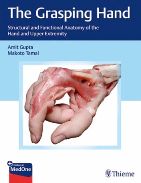
Anatomical guide leverages exceptional dissection images to elucidate the biomechanics of the hand and
upper extremity
The hand is a unique instrument that executes the commands of the brain and expresses the nuances of the
mind. The Grasping Hand: Structural and Functional Anatomy of the Hand and Upper Extremity by Amit Gupta
and Makoto Tamai is a state-of-the-art book that details the functions of the hand to feel, receive,
gather, collect and hold, as well as the complex role that the whole upper extremity plays in enabling
these actions. The anatomical structures intrinsic to these functions are detailed through illuminating
cadaveric dissections and succinct text.
Organized in 5 sections and 38 chapters, the book begins with a chapter detailing the intriguing history of
hand anatomy, followed by a section encompassing the structural and functional fundamentals. The third
section covers general anatomy and function, with discussions of the nerves and vascularity of the upper
extremity, as well as the brachial plexus. The fourth section features 26 anatomically organized chapters
from the shoulder to the fingertip with anatomical and functional insights on the joints, fascia and
retinacula, interosseus membrane, tendons and more. The single chapter comprising the final section covers
imaging and anatomy.
Key Highlights
anatomy and biomechanics
visual insights about underlying tissues and structures
and pathology
This practical resource is ideal for reviewing anatomy and biomechanics prior to performing hand, wrist,
arm, elbow, and shoulder surgery, making it essential reading for orthopaedic surgeons, fellows, and hand
specialists. This book is also useful for students of human anatomy, physical and occupational therapists,
medical students, and anyone interested in upper extremity anatomy and function.
This book includes complimentary access to a digital copy on https://medone.thieme.com.
Section I Prolegomena
1 The Story of Hand Anatomy
Section II Structural and Functional Fundamentals
2 Structural and Functional Anatomy of the Hand
2.1 Introduction
2.2 Prehension
2.3 Surface Anatomy
2.4 Structure
2.4.1 The Rays of the Hand
2.4.2 The Fixed and Mobile Elements
2.4.3 The Arches of the Hand
2.4.4 The Fibrous Skeleton
2.5 Muscles and Tendons
2.5.1 Extensor Tendons
2.5.2 Flexor Tendons
2.5.3 Interosseous Muscles
2.5.4 Lumbrical Muscles
2.5.5 The Intrinsic Muscles of the Thumb
2.5.6 Hypothenar Muscles
2.6 Movements of the Hand
2.7 Sensation and Proprioception
2.8 Control of Digital Motion
2.9 Grasp
2.10 Conclusion
3 Sense and Proprioception
3.1 Sensations
3.1.1 The Somatosensory Unit
3.2 Biomechanics of Proprioception
4 Joint Senses and Proprioception
4.1 Introduction
4.2 Innervation of Joints in the Human Hand
4.2.1 Wrist
4.2.2 Finger Joints
4.2.3 Thumb Trapeziometacarpal Joint
4.3 The Joint Senses and Proprioception
4.4 Conscious Joint Senses
4.5 Unconscious Joint Sense
5 The Hand and the Brain
5.1 Introduction
5.2 Motor System
5.3 Somatosensory System
5.4 Plasticity in the Somatosensory and Motor Systems
6 Structure and Function of Muscles
6.1 Overview
6.2 Muscle Architecture
6.2.1 PCSA and Fiber Length
6.2.2 Mechanics of Joint Motion
6.3 Molecular Anatomy
6.3.1 Active Tension
6.3.2 Passive Tension
7 Ultrastructure of Bones and Joints
7.1 Bone
7.1.1 Subchondral Bone Plate
7.1.2 Corner Zones
7.1.3 Physeal Scar
7.1.4 Trabecular Vault
7.1.5 Pillars
7.1.6 Diaphysis
7.1.7 Capsule
8 The Blood Vessels and Microcirculation
8.1 Vascular Supply to the Hand from the Base to the Fingertips
8.2 Differences between Large, Medium-Sized, and Small Arteries and Veins and the Vascular Network of the
Microcirculation
8.3 Outline of Anatomy and Physiology of Microcirculation for Metabolite Exchange
Section III General Anatomy and Function
9 Nerves of the Upper Extremity
9.1 Supraclavicular Branches of the Brachial Plexus
9.1.1 The Median Nerve
10 The Brachial Plexus
10.1 Introduction
10.2 Neural Anatomy
10.3 Vascular Anatomy
10.4 Lymphatic Anatomy
10.5 Posterior Triangle of the Neck
10.6 Exposure of the Brachial Plexus
10.7 Case Example 1: Adult Injury
10.8 Case Example 2: Obstetrical Injury
11 Vascular Anatomy of the Upper Extremity
11.1 Vascular Anatomy of the Upper Extremity
11.2 Axillary Artery
11.3 Brachial Artery
11.4 Ulnar Artery
11.5 Radial Artery
11.6 Veins of the Upper Extremity
11.6.1 The Superficial Veins of the Upper Extremity
11.7 The Deep Veins of the Upper Extremity
11.8 Lymphatic Vessels of the Upper Extremity
Section IV Regional Anatomy and Function
12 The Shoulder Joint
12.1 Introduction
12.1.1 Clavicle and Sternoclavicular Joint
12.2 Acromioclavicular Joint
12.3 Glenohumeral Joint
12.4 Glenoid, Labrum, and Capsule
12.5 Rotator Cuff
13 The Anatomy of the Arm
13.1 Humerus
13.2 Cutaneous Innervations of the Arm
13.3 Muscles of the Arm
13.4 Nerves of the Arm
13.5 Musculocutaneous Nerve (C5–C6)
13.6 Median Nerve in the Arm (C6–T1)
13.7 Ulnar Nerve in the Arm (C8–T1)
13.8 Radial Nerve in the Arm (C5–T1)
13.9 Arteries of the Arm
13.9.1 The Brachial Artery
13.10 Superficial Veins of the Arm
13.11 Summary
13.12 Surgical Approaches
13.13 Anterior Approach to the Humerus
13.14 Anterolateral Approach to the Humerus
13.15 Posterior Approaches to the Humerus
13.16 Extended Posterolateral Approach
14 The Elbow Joint
14.1 The Confluent Layered Anatomy of the Elbow
14.2 Articular Anatomy
14.3 Effect of Elbow Joint Morphology on Elbow Alignment
14.4 Structures That Provide for Varus/Valgus Stability
14.5 Structures That Resist Posterolateral Elbow Dislocation
14.5.1 Posteromedial Anatomy
14.5.2 Medial Anatomy
14.6 Deep Medial Anatomy
14.6.1 Anterior Anatomy
14.7 Distal Biceps Tendon
14.7.1 Lateral Anatomy
14.8 Lateral Collateral Ligament Complex
14.8.1 Posterior Anatomy
15 The Forearm Fascia and Retinacula
15.1 General Considerations for Fascia
15.2 The Fascial System of the Forearm
15.2.1 Retinacula System
15.2.2 Lacertus Fibrosus
15.3 Conclusion
16 Anatomy of the Forearm
16.1 Osteology
16.2 Myology
16.2.1 Volar
16.2.2 Dorsal
16.3 Arteries
16.4 Veins
16.5 Nerves
16.5.1 Cutaneous Nerves of the Forearm
16.5.2 Deep Nerves of the Forearm
16.6 Lymphatics
17 The Interosseous Membrane
17.1 Interosseous Membrane Anatomy
17.2 Kinematics
17.3 Biochemistry and Biomechanics
18 The Carpal Tunnel
18.1 Introduction
18.2 Anatomy
18.3 Borders
18.4 Contents
18.5 Surgical Anatomy
19 The Hypothenar Area: Anatomy of the Ulnar Carpal Tunnel
19.1 Anatomy of the Ulnar Carpal Tunnel
19.1.1 Guyon’s Canal
19.1.2 The Pisohamate Tunnel
19.1.3 The Opponens Tunnel
19.1.4 Anatomical Zones of the Ulnar Carpal Tunnel
19.2 Muscle Anatomy
19.2.1 Palmaris Brevis
19.2.2 Abductor Digiti Minimi
19.2.3 Flexor Digiti Minimi Brevis
19.2.4 Opponens Digiti Minimi
19.3 Nerve Anatomy
19.3.1 The Arborization of the Ulnar Nerve at the Hypothenar Area
19.3.2 The Branching Patterns of the Motor Branch to the ADM
19.3.3 The Deep Branch of the Ulnar Nerve
19.3.4 The Superficial Branch of the Ulnar Nerve
19.3.5 Other Communicating Branches
19.4 Vascular Anatomy
19.4.1 Anatomic Variations of the Ulnar Artery and Its Branches
20 Anatomy of the Wrist Joint
20.1 Bones of the Wrist
20.1.1 Extensor Retinaculum
20.1.2 Flexor Retinaculum
20.1.3 Ligaments of the Wrist
20.2 Extrinsic Ligaments
20.3 Radioscaphocapitate Ligament
20.4 Long Radiolunate Ligament
20.5 Radioscapholunate Ligament
20.6 Short Radiolunate Ligament
20.7 Dorsal Radiocarpal Ligament
20.8 Intrinsic Ligaments
20.9 Dorsal Intercarpal Ligament
20.10 Scapholunate Interosseous Ligament
20.11 Lunotriquetral Interosseous Ligament
20.12 Scaphotriquetral Ligament
20.13 Scaphotrapeziotrapezoidal Ligament
20.14 Soft-Tissue Attachments of the Pisiform
21 Vascularity of the Distal Radius and Carpus
21.1 Introduction
21.2 Vascular Anatomy of the Distal Radius
21.2.1 Extraosseous Vascularity
21.2.2 Intraosseous Vascularity
21.3 Vascular Anatomy of the Carpus
21.3.1 Extraosseous Vascularity
21.3.2 Intraosseous Vascularity
21.4 Vascularized Bone Grafts
22 Interosseous Vascularity of the Carpus
22.1 Introduction
22.2 The Lunate
22.3 Capitate
22.4 Scaphoid
23 Function of the Wrist Joint
23.1 Introduction
23.2 Wrist Kinematics
23.2.1 Flexion–Extension
23.2.2 Radial–Ulnar Deviation
23.2.3 “Dart-Throwing” Motion
23.3 Wrist Kinetics
23.3.1 Magnitude and Distribution of Forces across the Wrist
23.3.2 Primary Ligament Stabilization of the Carpus
23.3.3 Secondary Neuromuscular Stabilization of the Carpus
24 Anatomy of the Distal Radioulnar Joint
24.1 The Distal Radioulnar Joint (The MOBILE DRUJ)
24.2 The Extensor Retinaculum
24.3 The Triangular Fibrocartilage Complex (TFCC)
24.4 Discussion
25 Function of the Distal Radioulnar Joint
26 Hand Fascia, Retinacula, and Microvacuoles
26.1 Channelling of Structures in Transit Between Forearm and Digits
26.2 Restraint of Unwanted Motion
26.3 Transmission of Loads
26.4 Anchorage
26.5 Binding Role
26.6 Limiting or Tethering Role
26.7 Framework for Muscle Origins and Insertions
26.8 Vascular Protection and Pumping Action
26.9 Lubricating Role
26.9.1 The Microvacuolar System
26.9.2 Flexor Tendon Sheaths
27 Thumb
27.1 Osteology
27.2 Myology
27.3 Arteries
27.4 Veins
27.5 Nerves
28 The Flexor Tendons and the Flexor Sheath
28.1 Flexor Tendons
28.1.1 Flexor Carpi Radialis
28.1.2 Palmaris Longus
28.1.3 Flexor Carpi Ulnaris
28.1.4 Flexor Digitorum Superficialis/Sublimis
28.1.5 Flexor Digitorum Profundus
28.1.6 Flexor Pollicis Longus
28.2 Flexor Sheath
28.2.1 Synovial (Membranous) Component
28.2.2 Retinacular (Pulley) Component
29 The Extensor Tendons
29.1 Introduction
29.2 The Extensors Proximal to the Fingers
29.2.1 Extensor Retinaculum
29.2.2 Extensors of the Wrist
29.2.3 Extensors of the Thumb
29.2.4 Extensors of the Finger Metacarpophalangeal Joints
29.3 Extensors of the Fingers
29.3.1 Extrinsic Extensors of the Fingers and the Retinacular System
29.3.2 Intrinsic Extensors of the Fingers
29.4 Involvement of the Extensors in Control of Flexion of the Finger
29.5 Involvement of the Extensors in Control of Extension of the Finger
29.6 Conclusion
30 The Interossei
30.1 Anatomy and Biomechanics
30.2 Function
30.3 Summary
31 Lumbricals
31.1 Introduction
31.2 Detailed Anatomy
31.2.1 Origin
31.2.2 Insertion
31.2.3 Mechanics
31.3 Nerve Supply
31.4 Function
31.5 Pathologic Manifestations
31.5.1 Extensor Lacerations
31.5.2 Lumbrical Plus
32 Compartments of the Hand
32.1 Introduction
32.1.1 Thenar Compartment
32.1.2 Adductor Compartment
32.1.3 Interossei
32.1.4 Hypothenar Compartment
32.1.5 Carpal Tunnel
32.1.6 Finger
32.2 Compartment Monitoring
32.3 Compartment Release
32.4 Conclusion
33 Hand Spaces
33.1 Introduction
33.2 Perionychium/Pulp Space
33.3 Superficial Spaces
33.3.1 Subcutaneous Dorsal and Palmar Digital Spaces
33.3.2 Superficial Dorsal Hand Spaces
33.3.3 Palmar Subcutaneous Space
33.3.4 Interdigital Web Spaces
33.4 Synovial Spaces
33.4.1 Extensor Tendon Sheaths
33.4.2 Flexor Tendon Sheaths
33.4.3 Radial and Ulnar Bursae
33.5 Deep Potential Spaces
33.5.1 Space of Parona
33.5.2 Thenar Space
33.5.3 Midpalmar Space
33.5.4 Lumbrical Spaces
33.5.5 Hypothenar Space
34 The Carpometacarpal Joints
34.1 Introduction
34.2 Thumb Carpometacarpal Joint
34.3 Second Carpometacarpal Joint
34.4 Third Carpometacarpal Joint
34.5 Fourth Carpometacarpal Joint
34.6 Fifth Carpometacarpal Joint
34.7 Summary
35 The Metacarpophalangeal Joints
35.1 Surface Anatomy of Metacarpophalangeal Joint
35.1.1 Skin and Integument
35.1.2 Osteology
35.1.3 Capsule, Collateral Ligaments, and Volar Plate
35.1.4 Deep Transverse Metacarpal Ligament (Inter Palmar Plate Ligament) and the Metacarpal Transverse Arch
35.1.5 Extensor Apparatus
35.1.6 Flexor Tendons and Pulleys
35.2 Neurovascular Anatomy
35.3 Thumb Metacarpophalangeal Joint Surface Anatomy
35.3.1 Osteology
35.3.2 Capsule and Collateral Ligaments
35.3.3 Volar Plate and Sesamoids
35.3.4 Extensor Apparatus
35.3.5 Flexor Tendons and Pulleys
35.4 Neurovascular Anatomy
36 The Interphalangeal Joint
36.1 Introduction
36.2 Proximal Interphalangeal Joint
36.2.1 Bony Anatomy
36.2.2 Stabilizing Restraints
36.2.3 Vascular and Neurologic Structures
36.2.4 Surface Anatomy
36.3 The Distal Interphalangeal Joint
36.3.1 Bony Anatomy
36.3.2 Synovial Membrane
36.3.3 Stabilizing Restraints
36.3.4 Vascular and Neurologic Structures
36.3.5 Surface Anatomy
36.4 Biomechanics of Digital Motion
36.5 Summary
37 The Nail and Finger Pulp
37.1 Introduction
37.2 Embryology
37.3 Anatomy
37.3.1 Surface Anatomy
37.2.2 Longitudinal and Cross-sections of the Fingertip
37.2.3 Blood Supply to the Fingertip
37.2.4 Nerve Supply
Section V Epilegomena
38 Imaging and Anatomy
38.1 Introduction
38.2 Imaging Approach to Wrist Pathology
38.3 Anatomical Considerations
Index


