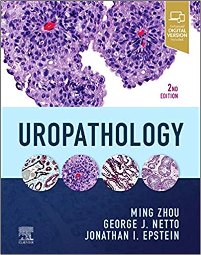
Designed for quick reference and efficient accurate sign-outs Uropathology 2nd Edition provides superbly illustrated expert
guidance in a time-saving format. This updated volume in the High-Yield Pathology series is highly templated for ease of use
featuring bulleted text authoritative content and high-quality images that comprehensively cover both non-neoplastic and
neoplastic entities making it easy to recognize the classic manifestations of urologic diseases and quickly confirm your
diagnoses.
Key Features
Provides in-depth, bulleted outlines for each entity covering definition, anatomy, and pathology: histology,
immunohistochemistry, and differential diagnosis.
Features 1,600 high-quality illustrations that include gross, radiographic imaging, microscopic, immunohistochemical, and
special stains, providing a comprehensive visual summary of the typical features of each entity.
Includes expanded information on tumor staging expertly provided by Dr. Ming Zhou, primary author of the College of American
Pathologists cancer protocols.
Reflects recent guidelines and protocols including:
Restructured tumor classification and new entities
New and modified grading systems for GU cancers
New and modified staging criteria for all GU cancers
New recommendations for reporting
New IHC and molecular markers for diagnosis and prognosis for GU cancers
Enhanced eBook version included with purchase, which allows you to access all of the text, figures, and references from the
book on a variety of devices
Author Information
By Ming Zhou, MD, PhD, Dr. Charles T. Ashworth Professor of Pathology, Director of Anatomic Pathology, UT Southwestern
Medical
Center, Dallax, Texas ; George Netto, MD, Professor and Chair of Pathology, University of Alabama at Birmingham School of
Medicine, Birmingham, Alabama and Jonathan I Epstein, Reinhard Professor of Urologic Pathology, Director of Surgical
Pathology,
Department of Pathology, The Johns Hopkins Medical Institutions, Baltimore, Maryland


