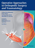
412 pp, 747 illustrations
Originally founded by three renowned orthopedic masters, Bauer, Kerschbaumer, and Poisel, this atlas has enjoyed a
longstanding reputation for the exceptional quality of its surgical, topographic anatomical illustrations. Considerable
advances in minimally invasive, endoscopic, and arthroscopic surgery during the last few decades necessitated this
updated edition.
Special Features
The addition of more than 40 new surgical procedures, including soft tissue preservation techniques used in trauma
surgery.
More than 700 color illustrations drawn directly from cadavers or documentation from the operating room.
The authors provide a unique operative guide, gleaned from years of evidence-based, clinical experience. From the
cervical spine to the foot and ankle, each part of the body is divided into detailed subsections. Multiple approaches
are included to treat common and rare musculoskeletal injuries, conditions, and diseases. The concise descriptions are
complemented by meticulously crafted, labelled anatomical drawings illustrating each step of the procedure, from the
skin incision to the targeted region to incision closure. Associated indications, patient positioning and preparation,
precautions, and dangers are also summarized for each approach.
This comprehensive, state-of-the-art atlas is an invaluable surgical resource for all orthopedic surgeons, residents,
and medical students.


