유방생검의 최신기법 - 진공보조 유방생검
- 저 자 :
- 역 자 : 박해린
- 출판사 : 가본의학
- ISBN(13) : 9788992006842
- 발행일 : 2010-09-02 / 1판 / 225 페이지
- 상품코드 : 22246
- 적립금: 1,080원

1 Senologic 소견의 문서화와 상관성 Documentation and Correlation of Senologic Findings 1
Renzo Brun del Re
1.1 도입 Introduction 1
1.2 Senometry 1
1.2.1 준비물 Material 1
1.3 임상 소견 표시하기 Mapping Clinical Findings 3
1.4 2 cm 보다 작은 유방 병변 위치 표시하기 Mapping Mammographic Lesions Smaller than 2 cm 3
1.4.1 유방촬영상에 나타난 병변에 대한 위치 표시 Mapping the Lesion on the Mammogram 3
1.4.2 유방촬영상의 수치를 유방 위로 옮기기 Transferring the Mammographic Dimensions onto the Breast7
1.5 수술 중의 senometry를 이용한 침 위치 결정법 Intraoperative Senometric Needle Localization 15
1.6 2 cm 보다 큰 유방촬영상의 유방병변의 도식화 Mapping Mammographic Lesions Larger than 2 cm 15
1.6.1 유방촬영상 나타난 병변 위치파악하기 Mapping the Lesion on the Mammogram 17
1.6.2 측정된 수치를 유방 위로 옮기기 Transferring Dimensions onto the Breast 18
1.7 초음파상 확인된 비 촉지성 병변 Nonpalpable Lesion Detected by Ultrasound 20
1.8 Senometry의 이점 Advantages of Senometry 21
References 22
2 대구경 진공보조 유방 생검과 절제 생검의 비교
Comparison of Large-Core Vacuum-Assisted Breast Biopsy and Excision Systems 23
Robin Wilson and Sanjay Kavia
2.1 서론 Introduction 23
2.2 대구경 중심 생검 시스템 : 개관 Large-Core Biopsy Systems: Overview 24
2.2.1 단일 대구경-중심 생검 시스템 Single Large-Core Biopsy System 24
2.2.2 진공-보조 유방 생검 시스템 Vacuum-Assisted Mammotomy Systems 26
2.3 적응과 한계 Indications and Limitations 36
2.3.1 한계점 Limitations 37
2.3.2 진단적 생검 Diagnostic Biopsy 37
2.3.3 치료적 절제술 Therapeutic Excision 39
2.3.4 이미지 유도하 생검 기술 Image-Guided Biopsy Technique 39
2.4 결론 Conclusions 40
References 40
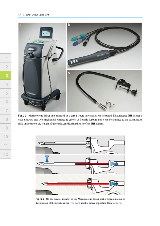
3 핸드 헬드 맘모톰을 이용한 초음파 유도하 진공 보조 유방 생검술
Sonographically Guided Vacuum-Assisted Breast Biopsy Using Handheld Mammotome 43
Luc Steyaert, Filip Van Kerkhove, and Jan W. Casselman
3.1 서론 Introduction 43
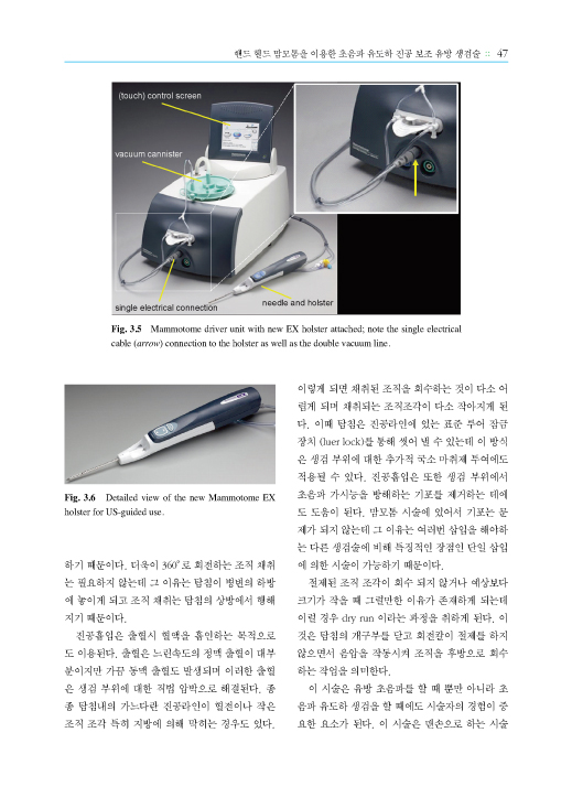
3.2 장비 Equipment 43
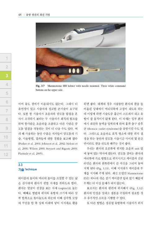
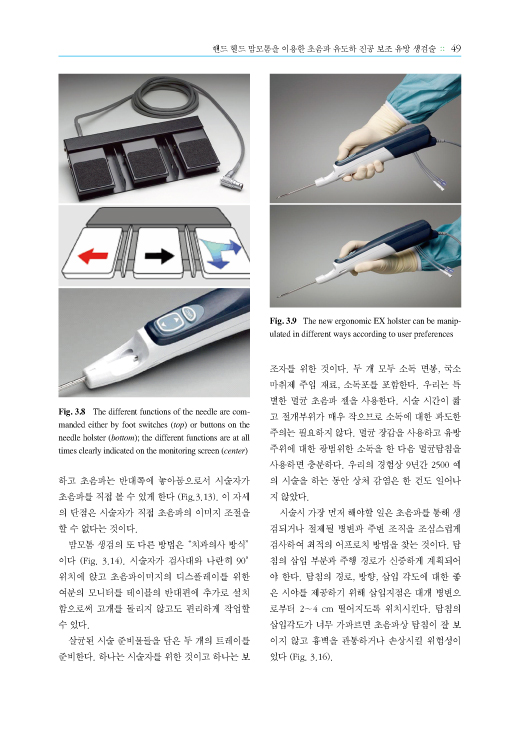
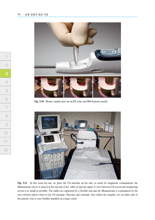
3.3 기술 Technique 48
3.4 적응증 Indications 67
3.4.1 양성 의심 혹은 불확실한 결절성 병변 Probably Benign or Indeterminate Nodular Lesions
68
3.4.2 매우 작은 크기의 의심 병변들 Very Small Suspicious Lesions 70
3.4.3 국소 감쇠 부분 Areas of Localized Attenuation 72
3.4.4 독립된, 복합 섬유 낭종성 부분들 Isolated, Complex Fibrocystic Areas 73
3.4.5 유두종 Papillomas 75
3.4.6 군집성 미세석회화 Clusters of Microcalcifications 76
3.4.7 어려운 위치에 있는 병변들 Lesions in Difficult Locations 79
3.4.8 이전 생검 또는 FNAC 상 비 결정적인 매우 단단한 병변들
Very Hard Lesions with Inconclusive Previous Biopsy or FNAC 82
3.4.9 불충분한 FNAC 또는 미세생검 결과 Inadequate FNAC or Microbiopsy Results 82
3.4.10 양성 병변의 제거 Removal of Benign Lesions 82
3.4.11 논의된 적응증들 : 방사상 반흔, 큰 낭종 내 병변들
Indications Discussed: Radial Scar, Large Intracystic Lesions 83
3.4.12 다른 적응증들 Other Indications 85
3.5 중심 검체 처리하기 Processing the Cores 85
3.6 탐침의 굵기 Needle Size 88
3.7 클립 위치시키기 Clip Placement 90
3.8 초음파 유도 Vs 유방촬영상 유도 US Versus Mammographic Guidance 90
3.9 결과 Results 92
References 93
4 Vacora 생검 시스템 TheVacora Biopsy System 97
R. Schulz-Wendtland
더보기
4.1 도입 Introduction 97
4.2 기술 Technique 97
4.3 적응증과 금기사항 Indications and Contraindications 98
4.3.1 초음파 유도하 경피적 진공 생검 US-Guided Transcutaneous Vacuum Biopsy 98
4.3.2 정위적 진공 생검 Stereotactic Vacuum Biopsy 101
4.3.3 MRI 유도하 진공 생검 MRI-Guided Vacuum Biopsy 101
4.4 부작용 Side Effects 101
4.5 결과 Results 101
4.6 한계 Limitations 102
4.7 실용적인 힌트 Practical Hints 102
References 102
5 정위적 유방 생검 Available Stereotactic Systems for Breast Biopsy 105
Ossi R. K쉉hli
5.1 도입 Introduction 105
5.1.1 엎드린 자세 방식 Prone Position Techniques 105
5.1.2 수직 시스템 Upright Systems 111
5.2 시스템의 비용 Cost of the Systems 112
5.3 시술의 비용 Cost of the Procedures 112
References 113
6 MRI 유도하 최소 침습적 유방 생검 MRI-Guided Minimally Invasive Breast Procedures 115
Harald Marcel Bonel
6.1 도입 : MR 맘모그래피의 역할 Introduction: Role of MR Mammography 115
6.1.1 적응증 Indications 116
6.1.2 기술과 실제적인 팁 Technique and Practical Tips 117
6.1.3 한계 Limitations 126
6.2 결론 Conclusion 127
References 128
7 유관내 종양에 대한 유관내시경 Ductoscopy of Intraductal Neoplasia of the Breast 129
Michael H웢erbein, Matthias Raubach, Y.Y. Dai, and Peter M. Schlag
7.1 도입 Introduction 129
7.2 유관내 유방 세포를 채취하기 위한 방법 Methods for Sampling Intraductal Breast Cells
129
7.2.1 유관내시경 기술 Technique of Ductoscopy 131
7.2.2 유관내시경 생검 Ductoscopic Biopsy 132
7.2.3 유두 분비물이 있는 여성에서의 유관내시경 Ductoscopy in Women with Nipple Discharge
132
7.2.4 유방암에서의 유관내시경 Ductoscopy in Breast Cancer 133
7.3 요약 Summary 134
References 134
8 최소 침습적 유방 생검 조직의 병리
Pathology of Breast Tissue Obtained in Minimally Invasive Biopsy Procedures 137
Gad Singer and Sylvia Stadlmann
8.1 도입 Introduction 137
8.2 최소 침습적 유방 생검 조직의 병리 Pathology of Breast Disease in Minimally Invasive Biopsies
137
8.3 양성 상피질환 Benign Epithelial Lesions 138
8.3.1 관주위 유방염 Periductal Mastitis 138
8.3.2 섬유낭종성 변화 Fibrocystic Change 139
8.3.3 경화성 선증 Sclerosing Adenosis 139
8.3.4 원주세포 병변 Columnar Cell Lesions 140
8.3.5 보통의 상피 증식 Usual-Type Epithelial Hyperplasia 141
8.3.6 소엽성 종양 Lobular Neoplasia 142
8.4 섬유상피성 병변 Fibroepithelial Lesions 142
8.4.1 섬유선종 Fibroadenoma 142
8.4.2 엽상 종양 Phyllodes Tumor 143
8.4.3 양성 유두상 병변 Benign Papillary Lesions 144
8.5 비침윤성 암 Malignant Noninvasive Lesions 145
8.6 침윤성 암 Invasive Carcinoma 146
8.7 유방암의 등급 결정 Grading of Breast Carcinoma 147
8.8 최소 침습적 생검에서의 예측 인자 Predictive Factors in MIBS 147
References 147
9 최소 침습적 유방 생검의 한계 Limitations of Minimally Invasive Breast Biopsy 149
Mathias K. Fehr
9.1 기술적인 실패 Technical Failures 149
9.2 최소 침습적 유방 생검에서 유방 병리의 과소평가
Underestimation of Breast Pathology on Minimally Invasive Breast Biopsy Specimens 150
References 155
10 유방 영상의 발전: 딜레마 혹은 진보? Advances in Breast Imaging: A Dilemma or Progress? 159
Daniel Fl쉜y, Michael W. Fuchsjaeger, Christian F. Weisman, and Thomas H. Helbich
10.1 도입 Introduction 159
10.2 초음파 Ultrasound 160
10.2.1 다면 표시 모드 Multiplanar Display Mode 161
10.2.2 함요 모드 영상 Niche Mode View 161
10.2.3 표면 모드 Surface Mode 161
10.2.4 투명 모드 Transparency Mode 161
10.2.5 정적 3차원 용적 대조영상 Static 3D Volume Contrast Imaging 162
10.2.6 4차원 용적 대조도영상 4D Volume Contrast Imaging 162
10.2.7 역전 모드 Inversion Mode 163
10.2.8 용적 계산 Volume Calculation 163
10.2.9 단층 초음파 영상 Tomographic Ultrasound Imaging 163
10.2.10 유리체 렌더링 Glass Body Rendering 164
10.2.11 강화 도플러, 색채 도플러, 그리고 고선명도 흐름
Power Doppler, Color Doppler, and High-Definition Flow 164
10.2.12 연장된 시야 기록 Extended View Documentation 165
10.2.13 결론 Conclusion 167
10.3 자기 공명 영상 Magnetic Resonance Imaging (MRI) 167
10.3.1 1.5-테슬라 시스템과 Gadopentate 1.5-Tesla Systems and Gadopentetate 167
10.3.2 3.0-테슬라 시스템 3.0-Tesla Systems 168
10.3.3 고분자 조영제 Macromolecular Contrast Agents 168
10.3.4 종양특이 조영제 Tumor-Specific Contrast Agents 170
10.3.5 기능적 유방 영상 기법 (분광법, 확산강조영상)
Functional Breast Imaging Techniques (Spectroscopy, Diffusion-Weighted Imaging) 170
10.4 양전자 방출 단층촬영 Positron Emission Tomography 172
10.4.1 세포 증식과 세포자멸사의 영상 Imaging of Cellular Proliferation andApoptosis 172
10.4.2 수용체 영상 Receptor Imaging 173
10.5 광학 영상 Optical Imaging 173
10.5.1 헤모글로빈 영상 Imaging of Hemoglobin 174
10.5.2 CTLM 장치 CTLM Device 174
10.5.3 외인성 조영제의 광학 영상 Optical Imaging of Extrinsic Contrast Agents 174
10.6 Electrical Impedance Scanning 176
10.6.1 표적 EIS와 TransScan TS2000 Targeted EIS with TransScanTS2000 176
10.6.2 T-Scan 2000ED와 Screening EIS 179
10.7 결론 Conclusion 179
References 180
11 비용-효과 분석 Cost-Benefit Analyses 183
Renzo Brun del Re and Regula E. Burki
11.1 도입 Introduction 183
11.2 유방촬영술의 빈도 Frequency of Mammography 183
11.3 소환률 Recall Rate 184
11.3.1 선별 검사에서의 소환율 Recall Rate in Screening Programs 184
11.3.2 선별 프로그램 외의 소환율 Recall Rate Outside of Screening Programs 185
11.4 추가 검사의 분포 Distribution of Further Investigations 185
11.4.1 외과적 절개 생검 Open (Surgical) Biopsy 185
11.4.2 절개 생검의 대체 Substitution of Open Biopsies 185
11.4.3 다른 진단적 기법의 대체 Substitution of Other Diagnostic Procedures 186
11.5 경향과 시나리오 Trends and Scenarios 187
11.6 비용의 비교 Comparison of Costs 187
11.6.1 비용과 절약 Costs and Savings 189
11.7 의사 결정자는 바뀌었다 Decision Makers Have Changed 190
11.8 결론 Conclusions 192
References 192
12 최근 데이터의 체계적 검토와 메타분석 Systematic Review and Meta-analysis of Recent Data 195
Renzo Brun del Re and Regula E. B웦ki
12.1 최소 침습적 유방 생검의 임상적 적절성에 대한 증거
Evidence for the Clinical Relevance of Minimally Invasive Breast Biopsy 195
12.1.1 고문헌의 체계적 검토 Older Systematic Reviews 195
12.2 문헌 조사 방법 Literature Search Methods 195
12.2.1 수행에 대한 증거 Evidence on Performance 195
12.2.2 위험성과 안정성에 대한 근거 Evidence on Risks and Safety 197
12.2.3 데이터의 발표와 분석 Presentation and Analysis of the Data 198
12.3 데이터의 발표와 분석 Presentation and Analysis of the Data 200
12.3.1 사용된 증거 Evidence Used 200
12.3.2 결과 Results 207
12.4 MIBB의 안정성 Safety of MIBB 212
12.4.1 절대표준으로서의 절개생검 Open Biopsy as Gold Standard 212
12.4.2 14-G 중심 생검과 11-G 진공 보조 생검 14-G Core Biopsies and 11 -G Vacuum-Assisted Biopsies
212
12.4.3 MIBB의 안정성에 대한 결론 Conclusions on the Safety of MIBB 215
12.5 삶의 질 Quality of Life (HRQoL) 215
12.6 발표된 MIBB 데이터에 대한 요약과 토론 Summary and Discussion of the MIBB Data Presented
217
12.7 결론 Conclusions 220
References 222