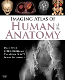
Softcover - 250 pages. International Edition.
Imaging Atlas of Human Anatomy, 4th Edition provides a solid foundation for understanding human anatomy. Jamie Weir,
Peter Abrahams, Jonathan D. Spratt, and Lonie Salkowski offer a complete and 3-dimensional view of the structures and
relationships within the body through a variety of imaging modalities. Over 60% new images-showing cross-sectional
views in CT and MRI, nuclear medicine imaging, and more-along with revised legends and labels ensure that you have the
best and most up-to-date visual resource. In addition, you’ll get online access to over 30 pathology tutorials linking
to additional images for even more complete coverage than ever before. In print and online, this atlas will widen your
applied and clinical knowledge of human anatomy.
Table of Contents
Preface to the Fourth edition
Preface to the First edition
Acknowledgements and Dedication
Introduction
1. Head, neck and brain
2. Vertebral column and spinal cord
3. Upper limb
4. Thorax
5. Abdomen and pelvis - Cross sectional
6. Abdomen and pelvis - Non cross sectional
7. Lower limb
8. Nuclear medicine
Index


