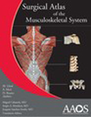
Containing over 960 beautiful anatomic images and full color photographs, this English edition of the original Manual y
Atlas Fotografico de Anatomia del Aparato Locomotor by Llusa, Meri, and Ruano, is a superb visual presentation of
musculoskeletal anatomy.
Preparing for MOC™ or primary certification? Use this excellent reference for a thorough review of musculoskeletal
anatomy.
Follow the images and amazing anatomic photographs as they are presented alongside corresponding MRIs or CT scans,
creating a multiplanar presentation with views of the anatomic areas in the coronal, frontal, and sagittal planes.
Scores of beautiful line drawings summarize the concepts and create a visual interconnectivity between anatomic
preparations, anatomic cuts, and images, demonstrating how different areas work together.
Included with the book is a DVD containing all of the figures and images from the book, for download and use in
presentations or for teaching.
차 례
Section I: General Information
Chapter 1: Terminology
Chapter 2: Osteology
Chapter 3: Arthrology
Chapter 4: Myology
Chapter 5: Neurology
Chapter 6: Angiology
Section II: Scapular Girdle and Upper Limb
Chapter 7: Anatomic Regions
Chapter 8: Osteology
Chapter 9: Arthrology
Chapter 10: Myology
Chapter 11: Neurology
Chapter 12: Angiology
Section III: Head and Trunk
Chapter 13: Topographic Regions and Surface Anatomy
Chapter 14: Osteology
Chapter 15: Arthrology
Chapter 16: Myology
Chapter 17: Neurology
Chapter 18: Angiology
Section IV: Pelvic Girdle and Lower Limb
Chapter 19: Topographic Regions and Surface Anatomy
Chapter 20: Osteology
Chapter 21: Arthrology
Chapter 22: Myology
Chapter 23: Neurology
Chapter 24: Angiology


