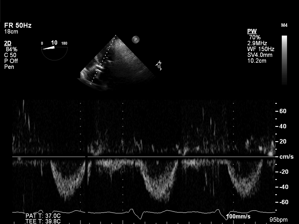
Figure 20.9F: Transesophageal echocardiographic deep transgastric view with still-frame of pulsed-wave Doppler of the left ventricular outflow tract in the same patient showing marked reduction in velocity time integral and therefore left ventricular stroke volume.
Movie 20.9E
|
Figure 20.9F
|
Figure 20.9G
