
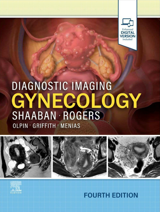
Diagnostic Imaging: Gynecology
- 저 자 : Shaaban
- 역 자 :
- 출판사 : Elsevier
- ISBN(13) : 9780443380396
- 발행일 : 2025-11-05 / 4판 / 944 페이지
- 상품코드 : 29949
- 적립금: 9,900원
- 정가: $357.99
Covering the entire spectrum of this fast-changing field, the fourth edition of Diagnostic Imaging: Gynecology is
an invaluable resource for general radiologists, specialized radiologists, gynecologists,
and trainees―anyone who requires an easily accessible, highly visual reference on today’s gynecologic imaging.
Drs. Akram M. Shaaban, Douglas Rogers, and their team of highly regarded experts provide up-to-date information on recent advances in technology and the understanding of pathologic entities to help you make informed decisions at the point of care.
The text is image-rich, with succinct bullets that quickly convey details, and includes the latest literature references, making it a useful learning tool as well as a handy reference for daily practice.
and their cell origins throughout
cell tumors (PEComas), and the newly described accessory cavitating uterine mass (ACUM)
(FIGO) and American Joint Committee on Cancer (AJCC) TNM
including radiology images, full-color medical illustrations, clinical photographs, and gross pathology and histology images, and includes all relevant modalities: Ultrasound (with 3D), sonohysterography, hysterosalpingography, MR, PET/CT, and gross pathology imagery
point of care
SECTION 1: TECHNIQUES
PELVIS
SECTION 2: UTERUS
INTRODUCTION AND OVERVIEW
AGE-RELATED CHANGES
CONGENITAL
INFLAMMATION/INFECTION
BENIGN NEOPLASMS
Endometrium
Myometrium
MALIGNANT NEOPLASMS
Endometrium
Myometrium
VASCULAR
TREATMENT-RELATED CONDITIONS
ADENOMYOSIS
SECTION 3: CERVIX
INTRODUCTION AND OVERVIEW
BENIGN NEOPLASMS
MALIGNANT NEOPLASMS
TREATMENT-RELATED CONDITIONS
MISCELLANEOUS
SECTION 4: VAGINA AND VULVA
INTRODUCTION AND OVERVIEW
CONGENITAL
BENIGN NEOPLASMS
MALIGNANT NEOPLASMS
LOWER GENITAL CYSTS
MISCELLANEOUS
SECTION 5: ADNEXA
INTRODUCTION AND OVERVIEW
PHYSIOLOGIC AND AGE-RELATED OVARIAN CHANGES
HORMONE-INDUCED OVARIAN CHANGES
INFLAMMATION/INFECTION
NEOPLASMS
Epithelial
Germ Cell
Sex Cord-Stromal
Miscellaneous
NONNEOPLASTIC ADNEXAL LESIONS
VASCULAR
ADNEXAL TORSION
SECTION 6: MULTIORGAN DISORDERS
INFLAMMATION/INFECTION
MALIGNANT NEOPLASMS
ABNORMAL SEXUAL DEVELOPMENT
SECTION 7: PELVIC FLOOR
ANATOMY
TECHNIQUE
PATHOLOGY